Tiếng Việt
Overview of dental cysts
Dental cysts are essentially epithelial cysts of the jawbone, covering part or all of the tooth crown or root. Most dental cysts develop from a decayed tooth or some reason that causes inflammation or infection of the tooth crown or root. When a part of the tooth is decayed or necrotic for some reason, it will release toxins back, making the inflammation worse. These areas of inflammation continue to stimulate the necrosis process, causing the Malassez epithelium around the dental ligament to be destroyed, creating dental cysts. The larger the cysts, the more they compress the tooth bone and release toxins that cause stronger tooth loss. This causes the bone to gradually wear away and become brittle.
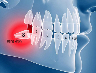
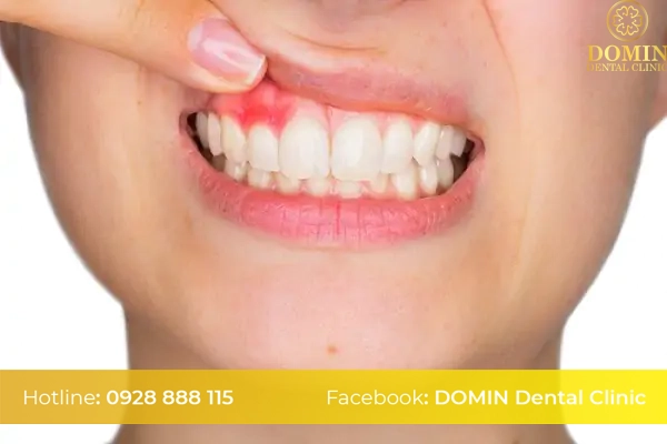
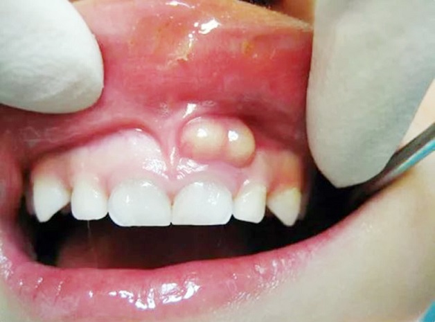
Dental cysts often develop silently and do not have any typical symptoms. Patients only feel pain when the cyst grows too large, swelling and toothache, showing signs of loosening. Dental cysts most commonly form at the location of the upper incisors. In addition, these dental cysts can form in any other location in the jaw.

Symptoms of this disease
Usually, dental cysts are difficult to detect. However, in some cases, dental cysts still manifest through symptoms such as:
– Slight discoloration of the teeth, most clearly observed at the root area. This can be considered an early warning sign of a dental cyst.
– When it reaches a severe stage, the jawbone swells, causing facial deformities. However, many people are subjective because this sign is not accompanied by pain, so they often ignore it.
– When pressing or touching the swollen area, it will feel hard (with small cyst size) and soft, indented feeling, meaning the cyst has grown in size.
– Causes loose teeth in surrounding areas.
– In children, there is a phenomenon of baby teeth not falling out even though the time for tooth replacement is past.
Are dental cysts dangerous?
Although the initial symptoms are often unclear and most people are unaware of the existence of dental cysts, this is a dangerous oral disease because it causes serious health effects.
– Dental cysts destroy tissue cells around the tooth tip. Gradually, when the tooth tip disappears, the inflammatory agents will progress to deep inflammation in the tooth root pulp, silently destroying the tooth root from the inside.
– Dental cysts cause local bone loss and are the cause of bone loss in neighboring teeth. If not treated promptly, it will lead to mass tooth loss.
– Dental cysts hinder the natural function of the jaw. When the jaw bone is reduced, chewing function is affected and the face is deformed.
When is dental cyst surgery needed?
When X-raying the patient’s dental cyst, the image shows a round or oval radiolucent area attached to a dead root. Accompanying symptoms are:
– The tooth corresponding to the cyst has pulp death and a large cavity.
– The surrounding teeth are often tilted, and if severe, can even be displaced.
– The ligaments around the teeth begin to stretch.
– There is a phenomenon of adjacent tooth root resorption due to long-term progression of the dental cyst.
– Cysts in the maxilla grow rapidly, can expand in many directions and become atypical.
– The cyst boundaries become unclear because of inflammatory vasodilation and surrounding bone resorption.

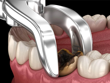
Surgical procedure
Before surgery, the patient will have a dental X-ray to identify the affected teeth and assess their condition. In cases where the teeth are too loose and more than 1/3 of the root bone has been lost, they will be extracted. Conversely, if there is enough alveolar bone left and the part of the root that is to be cut is smaller than the part that falls into the cyst, the teeth can be retained. These teeth are usually treated endodontically before surgery.
The doctor will then determine the size of the cyst, the level of bone loss and contact with surrounding areas such as the maxillary sinus, nasal cavity, etc. to choose the appropriate treatment method.
The steps involved in performing dental cyst surgery include:
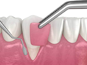
– Mucosal incision : the doctor will make a ruler-shaped or semicircular incision at the base of the corridor, without having to separate all the way to the top of the alveolar bone. Then, separate the mucosal periosteum from the bone using a specialized dissecting tool.
– Open the bone to expose the cyst : if the outer bone layer is thin, the cyst membrane will appear immediately after peeling off the mucosa. On the contrary, if the bone layer is thick, the doctor will use a drill or bone chisel to create a hole and then use forceps to widen it to almost the diameter of the cyst. This will avoid tearing the cyst membrane below. In case the bone window is not wide enough, the hole will quickly heal, fill in and the cyst will recur.
– Opening the cyst: the dentist uses specialized forceps to stretch the cyst membrane and then cuts an oval piece of the cyst membrane based on the shape of the bone window that has just been opened.
– Perform apical sectioning: the tooth is cut at the same level as the base of the cyst, then the remaining pieces of cyst membrane are used to cover the root sections, this process will help the wound heal quickly.
– End of surgery : the doctor will use a drill or bone file to smooth the bone edge, thoroughly wash the cyst cavity and then cover the mucosal periosteum flap into the tooth socket. Here, the doctor can create a self-hardening plastic button to press on the outside, this plastic button is often ground down based on the bone formation rate or use a removable denture base to cover the hole. At this stage, for large cysts, with a range of more than 3 teeth, the doctor will leave a part of the upper cyst membrane edge after cutting to then suture the edge with the mucosal edge, the remaining part of the cyst membrane is sutured outward with the mucosal flap. In the case of tooth extraction, the technique is similar, but when opening the cyst, pull the mucosal periosteum flap down to cover the cyst and the jaw bone surface as much as possible.
– Post-operative care : the patient is carefully cared for and instructed by the hospital doctor and nurses. The patient is instructed to rinse the mouth thoroughly to avoid leaving food in the bone socket. After 10-14 days, return for a check-up, grind down the plastic plug and remove it completely when the bottom of the cyst is full and close to the opening of the hole.
Prestigious address for dental cyst surgery
After 9 years of operation, Domin Dental has become a prestigious and high-quality dental care and treatment address for all customers across the country.
The team of doctors at the dental clinic are all experts with many years of experience, trained extensively abroad, and are at the forefront of updating and learning advanced technologies.
Not only focusing on the team of doctors and machines, but service is also one of the strengths at Domin Dental.
For direct consultation, readers can click here.


Frequently asked questions about dental cyst curettage
Usually the surgery only takes about 30 – 60 minutes. Since this is a minor surgery, the treatment time is not too long.
After surgery, if you follow your doctor’s advice and take good care of your wound, your recovery will be faster. Usually, after about 7 days, the wound at the surgical site will be able to heal normally.
The main treatment is surgery to completely remove the cyst inside. Therefore, most cases will not have a recurrence of the disease. However, there are some cases where the infection may recur due to poor wound care. Therefore, you should pay attention to oral health care in the postoperative period.